


 cho-planar imaging (EPI) is completely different from the imaging methods mentioned above. It is the fastest imaging sequence currently available, and unlike the other sequences discussed in this chapter, it does not use the spin-warp technique. However, in its latest implementation, it is conceptually very similar to spin warp.
EPI was proposed by Peter Mansfield in 1977 [⇒ Mansfield 1977]. It is based on the principle of a single excitation of the spins, followed by the rapid switching of a strong, high-performance gradient to form a series of gradient echoes, each of which is given a different degree of phase-encoding and thus can be reconstructed to form an image.
The phase gradient can be applied as a constant gradient, as in the original echo-planar scheme (Figure 08-07), or a series of small ‘blips’, each of which corresponds to one phase-encoding step [⇒ Johnson 1983].
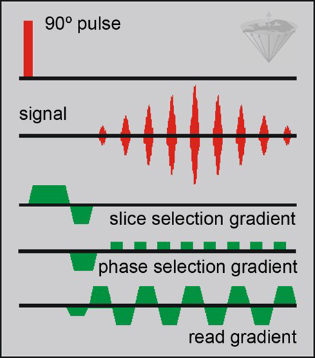
Figure 08-07:
An EPI pulse sequence (FID-based MBEST sequence). The example of this figure consists of 9 sampling periods. For a 64×128 image matrix, 64 sampling periods are necessary. During each sampling period, 128 points are sampled.
The k-space trajectory is a single sawtooth-pattern path (Figure 08-08).
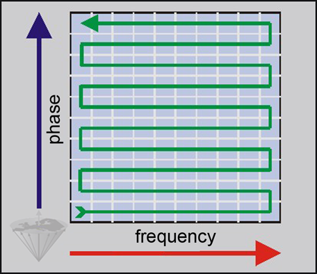
Figure 08-08:
The k-space trajectory of an echo-planar imaging experiment.
EPI is faster than FLASH, but FLASH has a higher spatial resolution.
One of the main problems with the original echo-planar sequence is the T2* dephasing of the signal during the scan.
One can reduce this effect by forming the EPI echo train about a spin echo, even though substantial T2* dephasing will remain at the start and end of the EPI sequence. To minimize such effects, very short scan times have to be achieved [⇒ Cohen 1991, ⇒ Pykett 1987].
However, as one reduces the sampling period, one also reduces the signal-to-noise ratio and increase the amplitude of the read gradient required to obtain a given resolution. For these reasons, single shot EPI tends to be limited to a maximum of a 128×256 matrix.
The true snapshot capability of echo planar is very attractive and has made EPI the fundamental tool to acquire most fMRI as well as tractography and thermometry data.
 Still, the problems associated with the technique mean that it could not be used for a number of possible other — clinical — applications. Developments in gradient and switching techniques have, however, partly overcome these problems, and single shot EPI is available on most new high field and ultrahigh field machines.
Single shot EPI still suffers from a chemical-shift artifact since the bandwidth per pixel in the phase-encoding direction is less than the chemical shift between water and fat. The applications for EPI are the same as those cited for snapshot FLASH, mostly diffusion imaging and functional imaging.
Multi shot EPI improves image quality tremendously. However, at ultrahigh fields drastically shortened T2* values cause a loss of signal for long TE gradient echo acquisitions and give rise to problems for echo-train based acquisition techniques such as EPI.
Another problem of EPI at high and ultrahigh fields are its hazards. There remains some uncertainty with respect to the safety of EPI since the very rapid switching of strong gradients generates electrical currents in the body which can stimulate peripheral or cardiac nerves. During the early years of EPI the developers in the USA and in Europe had to change their magnets from 2 T to 1 T to avoid physiological effects of fast oscillating field gradients [⇒ Budinger 1991, ⇒ Cohen 1990] (cf. Chapter 18).


|

|
|---|