


 ulk or macroscopic flow of blood in vessels and of cerebrospinal fluid adds still another parameter to image contrast in MR imaging. This kind of fluid motion is different from diffusion and perfusion and follows specific laws.
A number of contrast features of flow in MR imaging and in magnetic resonance angiography (MRA) are rather complicated. The explanations in this chapter give a general overview, without attempting to cover the complexity of the topic in detail (Figure 14-01).
Figure 14-01:
Checking the vessels: Will there be laminar or plug flow? Or, worse, will plaques create turbulent flow?
Flowing blood and CSF can appear bright or dark, depending on their velocity, direction and pattern of flow, and the pulse sequence used. In routine MR imaging normal flow effects can mimic pathology. Thus, understanding their influence upon image contrast is very important. This includes also knowledge of vascular anatomy and comprehension of vascular dynamics.
 Blood flowing through a small caliber vessel usually exhibits laminar flow (Figure 14-02). Because of shearing forces, the blood closest to the vessel wall flows slowest. Blood velocity increases towards the center. Laminar flow is the predominant kind of flow in the human body.
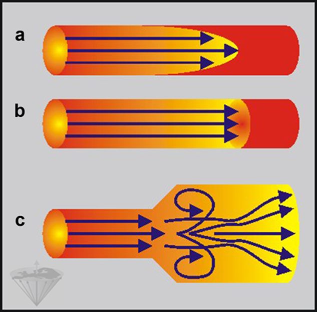
Figure 14-02:
(a) Laminar flow, (b) plug flow, and (c) laminar flow turning into vortex and turbulent flow after a vascular stenosis. Laminar flow is relatively slow, plug flow, as a special form of turbulent flow, is faster.
Blood flowing faster in larger caliber vessels develops turbulence, in particular at places where the vessel's diameter changes, e.g., after stenoses or in vessels with irregular lumen, and moves more randomly, which produces phase shifts among blood cells. The spins dephase and the blood signal intensity decreases.
Plug flow is a special case of turbulent flow with a flat flow profile; all fluid elements possess the same velocity. Laminar flow is relatively slow, whereas plug flow, as a special form of turbulent flow, is faster. Note that all flow patterns in Figure 14-02 consist of conceptual schemes.
In general, flow velocity differs from arteries to veins. Pulsatile flow in arteries, to a lesser extent also in veins, is cyclical and irregular, depending on systole and diastole of the heart. Thus, the appearance of the nature and velocity of the flow will depend on when during the cardiac cycle the image is taken. This kind of flow is often turbulent during parts of the cycle.


|

|
|---|