


 he spleen, pancreas, bone marrow, lymph nodes, adrenals, muscles, in particular the heart, as well as inflammations and specific tumors have been proposed as additional target regions for some contrast agents. None of these, nor a number of other agents not mentioned here, are available for clinical imaging.
Agents for contrast-enhanced MR angiography (i.e., ECF space and blood pool agents) are discussed in Chapter 14.
Dysprosium can be used as an intravenous negative contrast agent, as was shown with Dy-DTPA-BMA, a paramagnetic bulk susceptibility perfusion agent. It possesses a large magnetic moment and poor T1 relaxivity, despite its relationship with gadolinium and manganese.
It can be applied as T2-relaxation contrast agent at extremely high fields [⇒ Haraldseth 1993, ⇒ vander Elst 2002]. However, Dysprosium compounds are not used clinically.
Not all contrast agents are aimed at proton magnetic resonance imaging. One can also exploit magnetic properties of nuclei different from hydrogen, for instance fluorine. The feasibility of in vivo application of perfluorinated compounds has been demonstrated for lung ventilation and perfusion studies (Figure 20-40) [⇒ Rinck 1984].
Gadolinium-based aerosols, hyperpolarized gases, and oxygen were also proposed to be used as possible ventilation agents. In the case of hyperpolarized gases helium has been described as better suited than xenon [⇒ Albert, ⇒ Middleton]. However, special hardware is necessary: devices for production, storage, and installation of the hyperpolarized gases, as well as special coils and receivers for imaging. This makes ventilation imaging with these gases more difficult than comparable methods. The methods are only applied in a few clinical imaging facilities. An in-depth explanation of the technique was given by Roos et al. [⇒ Roos 2015].
Problems in abdominal MR imaging are created by motion artifacts from respiration, cardiac motion, peristalsis, and blood flow, as well as a general lack of contrast between feces- and fluid-filled or collapsed bowel loops and adjacent organs or pathological structures. Cardiac and respiratory gating, special software, faster imaging sequences, and the use of contrast agents have solved some of them.
A number of substances were proposed and studied during the last years in phantom, animal, and clinical trials. Among them were positive and negative contrast agents. Gadolinium-containing contrast agents belonged to the first group, while fluorine-containing compounds and magnetic particles are found in the second group (Figure 13-16) [⇒ Hamm 1992, ⇒ Rinck 1991]. For studies of the upper abdomen blueberry or pineapple juices are also useable.
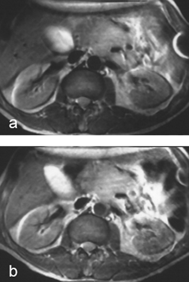
Figure 13-19:
Intermediately weighted images of the upper abdomen. Recurrent mesenteric tumor.
(a) plain; (b) after ingestion of a negative oral contrast agent. On the enhanced image, the contour of the tumor is well delineated from the neighboring liver and intestines.
Artifact created by ECG lead in the left abdominal wall.
The use of enteral contrast agents has been limited, and with today’s imaging techniques, in particular custom-tailored pulse sequences, most clinical questions can be answered without the application of oral or rectal agents.


|

|
|---|