


 he news of Lauterbur's invention traveled slowly although he presented it at a number of scientific meetings between 1972 and 1975. Some people listened, understood, and reacted. It's a tale of international conferences.
Here are some examples:
In April 1974, Lauterbur gave a talk at a conference in Raleigh, North Carolina. This conference was attended by Richard Ernst from Zurich (Figure 20-31), who realized that instead of Lauterbur's back-projection one could use switched magnetic field gradients in the time domain.
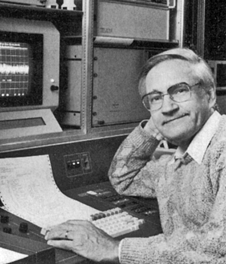
Figure 20-31:
Richard Ernst (1933-2021).
This led to the 1975 publication NMR Fourier Zeugmatography by Anil Kumar, Dieter Welti, and Richard Ernst [⇒ Kumar 1975], and to the universal reconstruction method for MR imaging today (Figure 20-32).
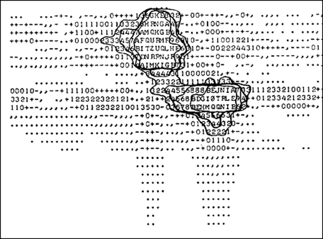
Figure 20-32:
One of the first 2D-FT MR images from the research group of Richard Ernst in Zurich, acquired by Anil Kumar in July 1974.
When Lauterbur presented his approach to NMR imaging at the International Society of Magnetic Resonance (ISMAR) meeting in January 1974 in Bombay, Raymond Andrew, William Moore, and Waldo Hinshaw from the University of Nottingham, England, were in the audience and took note.
 As a result, Hinshaw (Figure 20-33) developed his own approach to MR imaging with his sensitive point method [⇒ Hinshaw 1974; 1976].
 Figure 20-33: Waldo Hinshaw.
At this time, several research groups in Nottingham worked in parallel on similar topics. The first group comprised E. Raymond Andrew, Waldo S. Hinshaw, William S. Moore, Neil Holland, and Paul Bottomley, all of them major contributors to the development of MR imaging.
In 1977, Waldo Hinshaw, Paul Bottomley, and Neil Holland, succeeded with an image of the wrist [⇒ Hinshaw 1977]. Hinshaw later went to Harvard and then joined the group at the Technicare company, at that time the most advanced scientific group with a commercial and sound medical applications background.
More human thoracic and abdominal images by different groups and several novel techniques followed, and by 1978, Hugh Clow and Ian R. Young, working at the British company EMI, reported the first transverse NMR image through a human head [⇒ Clow 1978].
The second group in Nottinham included Peter Mansfield (Figure 20-34), Peter K. Grannell, Andrew Maudsley, Ian Pykett, and Peter Morris. They worked on studies of solid periodic objects, such as crystals.
 Figure 20-34: Peter Mansfield (1933-2017).
At a Colloque Ampère conference in Cracow, Poland, in September 1973, Mansfield was told about Lauterbur's imaging method after he and his collaborator Peter K. Grannell had presented a one-dimensional interferogram of camphor/cardboard samples (Figure 20-35) [⇒ Mansfield 1973].
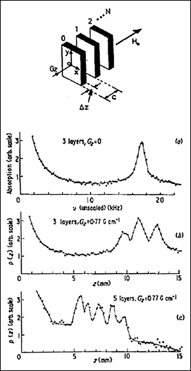
Figure 20-35:
Model of the one-dimensional lattice (camphor with cardboard spacers used for the experiment). Fourier cosine transforms of the transient response to a multiple pulse sequence from the layers of camphor. The result is a linear measurement in a single dimension, not an image.
(a) Three layers, no magnetic field gradient; (b) three layers, gradient of 0.77 G/cm; (c) five layers, same gradient.
For some time in 1974 a third research ‘team’ existed in Nottingham, consisting of Alan N. Garroway (Figure 20-36). He applied weak radiofrequency pulses in the presence of a field gradient in order to achieve spatial selectivity. Next door, Peter Mansfield and his postdoctoral students were developing a related method.
 Figure 20-36: Alan N. Garroway.
Garroway later joined the Mansfield group. A month before Garroway and Mansfield submitted their first imaging article to Journal of Physics in 1974 they applied together for a first patent [⇒ Garroway 1974]. Unfortunately, their method was unsuitable for practical application because it suffered from rapid loss of signal; the problem and its solution were explained by David Hoult from Oxford [⇒ Hoult 1977].
By 1975, Mansfield and Andrew A. Maudsley proposed a line technique which, in 1977, led to the first image of in vivo human anatomy, a single cross section through a finger. In 1978, using the same single-slice method, Mansfield presented his first image through the abdomen [⇒ Mansfield 1976; 1978].
 Figure 20-37: Roger Ordidge.
Echo-planar imaging (EPI), a real-time imaging technique, had been proposed by Mansfield's group in 1977, and the first crude images were shown by Mansfield and Ian Pykett in the same year. Roger Ordidge (Figure 20-37) presented the first EPI movie in 1981 [⇒ Ordidge 1981].
The breakthrough of EPI came with manifold improvements in many aspects of the associated methodology and instrumentation — from gradient power supply and gradient coil design to pulse sequence development, presented by Pykett and Rzedzian in 1987 [⇒ Pykett 1987]. However, it remains a niche technology in clinical MRI.
The group around John Mallard (1927-2021) at the University of Aberdeen also performed trailblazing research work. James Hutchison (1941-2018), a physicist, Margaret A. Foster, a biologist, and later William A. Edelstein (1944-2014), and their colleagues built their own whole-body MR imaging machine and developed the spin-warp technique.
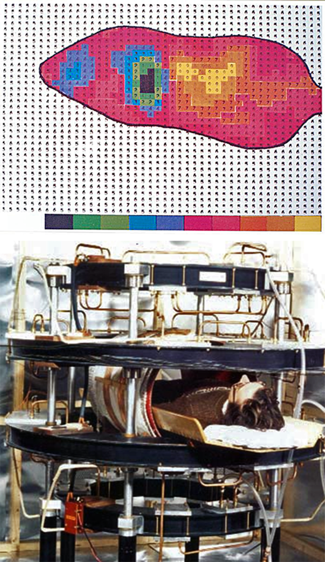
Figure 20-38:
(Top) The first image of a whole mouse was obtained in Aberdeen, Scotland, in March 1974.
(Bottom) The prototype whole-body MR machine in Aberdeen. James Hutchison (1941-2018) lies in the magnet.
They published the first image through the body of a mouse in 1974 which was followed by a human whole-body image in 1980 (Figure 20-38) [⇒ Hutchison 1974; ⇒ Edelstein 1980].
In the 1970s and 1980s Great Britain was a major contributor to the development of MRI equipment and software, but then a number of the researchers working in Britain went to the United States. It was a major brain-drain for British universities, but there was little money in the British university system.
An excellent eyewitness report by some leading British researchers and scientists about the British work was given in the transcripts of a meeting at the Wellcome Institute for the History of Medicine, London, in 1996 [⇒ Christie 1998].
Most of the British researchers stayed abroad, whereas many of the Continental Europeans who worked in the U.S.A. in the late 1970s and early 1980s returned to Europe. Some of the Europeans had performed quite impressive research in the United States.
Among them was Robert N. Muller who, in 1982, described off-resonance imaging, a technique later dubbed magnetization transfer imaging (Figure 20-39) [⇒ Muller 1983]. Peter A. Rinck et al. described the first in vivo fluorine lung images (Figure 20-40) [⇒ Rinck 1984].
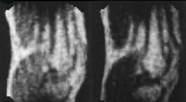
Figure 20-39:
Off-resonance images of a knee joint (1982).
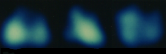
Figure 20-40:
19F ventilation imaging of a dog's lung; coronal and transverse images (1982).
Much of the research done at this time was not published, or only presented as abstract in the proceedings of scientific meetings because of the extremely rapid progress in the different research groups.
In vivo MR Spectroscopy. Actual in vivo NMR spectroscopy took off in Oxford from 1974, with the group of Rex E. Richards and George K. Radda. Among others, David Hoult and David G. Gadian belonged to this group [more details can be found at ⇒ Christie 1998].


|

|
|---|