


 inding a relevant use of this new technique was difficult, and medicine and biology stayed somewhere backstage although in vivo NMR with a medical background has its roots in the early and mid-1950s.
In 1955 Erik Odeblad (Figure 20-17) and Gunnar Lindström from Stockholm published their first NMR studies, including relaxation time measurements of living cells and excised animal tissue [⇒ Odeblad 1955].
Odeblad is the main pioneer in NMR in medicine and laid the foundations of NMR and MRI in biomedicine.
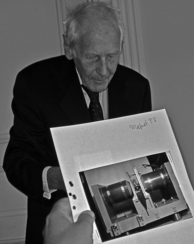
Figure 20-17:
Erik Odeblad (1922-2019), recalling his first NMR spectrometer after receiving the European Magnetic Resonance Award 2012 — 57 years after his first publication about relaxation times and NMR in medicine.
In 1952, while working at the University of California in Berkeley Odeblad met Felix Bloch in Stanford. He asked him whether he could use Bloch's NMR spectrometer to study human samples, but the response was negative: NMR was a tool for physicists, not for research into physiology, medicine, or biology.
Odeblad returned to Sweden — and got his own machine. Around the year 1950 Gunnar Lindström of the Nobel Institute of Physics in Stockholm had built a spectrometer. Odeblad adapted and used it for his pioneering biomedical NMR applications, in vivo and ex vivo. In December 1954, they submitted their first NMR results [⇒ Odeblad 1955] (Figure 20-18a).
They had found out that different tissues had distinct relaxation times, most likely due to water content but also to different bindings to lipids — a phenomenon that explains tissue contrast in MR imaging. Odeblad continued working on human fluids and tissues throughout the following decades and some sixty scientific papers on NMR in human tissues and secretions of mucous membranes followed between 1955 and 1968 [⇒ Odeblad 1957].
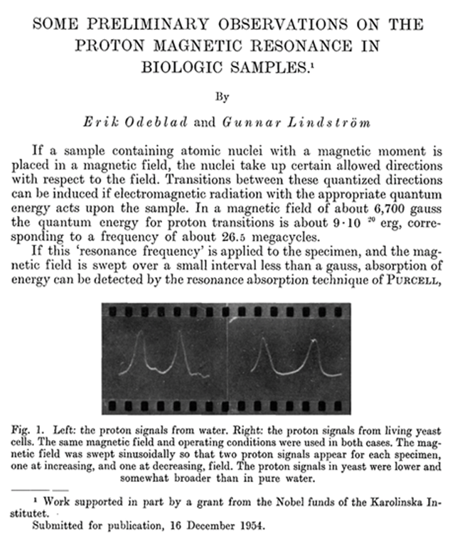
Figure 20-18a:
The first paper on biological and medical NMR: "Some preliminary observations on the proton magnetic resonance in biological samples", by Erik Odeblad and Gunnar Lindström, submitted for publication to Acta Radiologica (Stockholm) in December 1954, published in 1955.
The research for these publications was performed at the Department of Obstetrics and Gynecology at the Sabbatsberg Hospital, Karolinska Institute, and the Nobel Institute of Physics in Stockholm. It was partly supported by a grant given by the Nobel Funds.
He extended the range of possible applications further in the fields of obstetrics and gynecology (his first medical discipline used to be gynecology and he became world-known for his research in fertility) [⇒ Odeblad 1959]. He was sure that magnetic resonance would become an asset in medicine (Figure 20-18b).
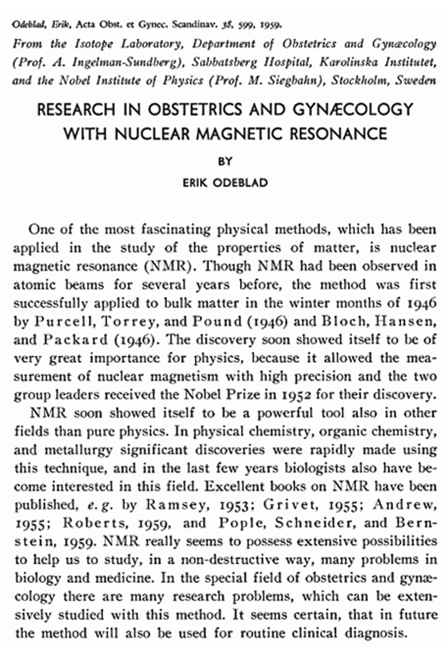
Figure 20-18b:
From Odeblad’s 1959 paper on magnetic resonance in obstretrics and gynecology: “NMR seems to possess extensive possibilities to help us study, in a non-destructive way, many problems in biology and medicine … It seems certain that in future the method will also be used for routine clinical diagnosis.”
Later he also tried to localize cancerous tissue in vivo, but did not succeed [⇒ Ingelman-Sundberg, Odeblad 1965]. In this paper the authors state: “… carcinoma of the uterus, which in all cases gave rise to significant RF absorption differences between contralateral symmetrical points. The results were fully confirmed at hysterectomy. By intrauterine RF scanning it appears, therefore, possible to determine the surface spread of an endometrial carcinoma.” However, it was done solely by RF, not by NMR.
Odeblad finally left Karolinska for, what is claimed, reasons of conscience related to his fertility research, and the mandatory introduction of abortion at Karolinska. He wrote a second PhD thesis in medical physics and became Professor of Medical Biophysics at the newly founded University of Umeå in northern Sweden.
A forgotten pioneer …
 Erik Odeblad's disregarded discovery …
 Soon others joined in this kind of research. In the late 1950s and early and mid 1960s the results of a very large amount of work on relaxation, diffusion, and chemical exchange of water in cells and tissues of all sorts appeared in the scientific literature.
Oleg Jardetzky and his collaborators performed sodium NMR studies in blood, plasma and red blood cells in 1956 [⇒ Jardetzky 1956]. T1- and T2-measurements of living frog skeletal muscle were published by Bratton in 1965 [⇒ Bratton 1965]. In 1967, Ligon reported the measurement of NMR relaxation of water in the arms of living human subjects [⇒ Ligon 1967].
Then, in the late 1960s and early 1970s research and dedicated science mushroomed in the field.
In the late 1960s, James Hutchison began working with magnetic resonance on in vivo electron spin resonance studies in mice at the University of Aberdeen in Scotland. Hazlewood added to the work on NMR relaxation time measurements by studying developing skeletal muscle tissue [⇒ Hazlewood 1969, 1971]. Cooke and Wien worked on similar topics [⇒ Cooke 1971]. Hansen focussed upon NMR studies of brain tissue [⇒ Hansen 1971].
The first animal whole-body spectrometer was built in 1968. Jackson and Langham working at the Los Alamos Scientific Laboratory of the University of Los Angeles obtained the first NMR signal ever from a whole living animal, an anesthetized rat (Figure 20-19) [⇒ Jackson 1968].
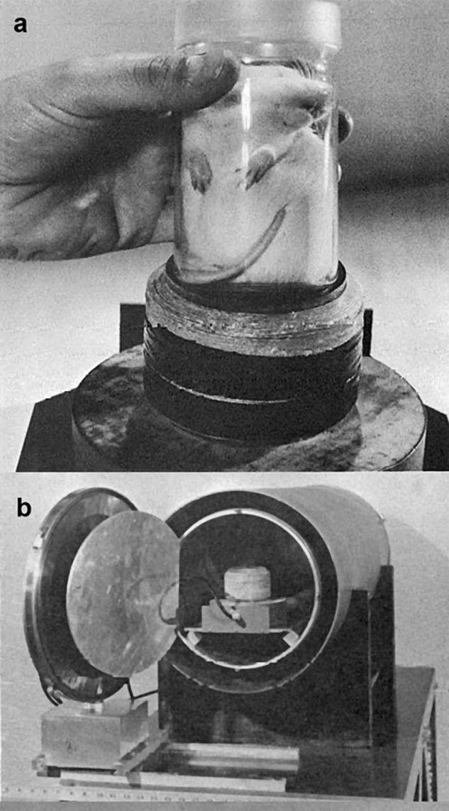
Figure 20-19:
The Los Alamos NMR spectrometer operating at 0.001 T (1968).
(a) A rat being inserted into the sample coil. (b) End view of the solenoid and magnetic shield after assembly with sample coil.
At this time medical doctors without prior background in NMR science joined in the research, curious about possible biological applications of relaxation times.
One of them was Raymond Damadian (1936-2022) at Downstate Medical Center in Brooklyn. He measured relaxation times of excised normal and cancerous rat tissue and stated that tumorous tissue had longer relaxation times than normal tissue [⇒ Damadian 1971].
There was a lack of controls in his measurements and, as it turned out quickly, it was a fallacious assumption.
Donald P. Hollis and his colleagues from Johns Hopkins University in Baltimore repeated Damadian’s studies — on the same pulsed NMR spectrometer Damadian had used and Paul C. Lauterbur had provided. Hollis reached conflicting, contrary results and was more cautious and critical in his scientific conclusions. There was no verification of Damadian’s claims that cancer pathology and their relaxation times possessed a numerical correlation [⇒ Hollis 1972, 1973].
Yet, Damadian promoted his findings as the ultimate technology to screen for (in Damadian's words: "to scan" — but not to image) cancer and patented the idea of a hypothetical relaxation time scanner similar to the Los Alamos spectrometer as “Apparatus and method for detecting cancer in tissue” (Figure 20-20c) [⇒ Damadian 1974].
In February 1973 Zenuemon Abe and his colleagues applied for a patent on a targeted NMR scanner [⇒ Abe 1973] (Figure 20-20b). They published this technique in 1974 [⇒ Tanaka 1974]. Damadian reported a similar technique in a publication two years later, dubbed 'field-focusing NMR (Fonar)' which contained an image composed of one-dimensional measurements: step-by-step scanned volume elements through a mouse [⇒ Damadian 1976]. However, his Fonar company that manufactured MRI equipment used the MR imaging method described by Paul Lauterbur. Neither Abe's nor the Fonar techniques were suitable for medical imaging.
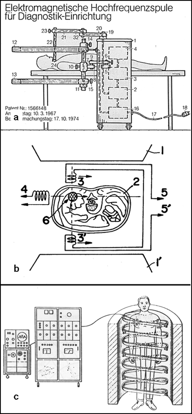
Figure 20-20:
As examples among others: Graphic designs of three patented though practically inapplicable magnetic field based diagnostic systems. All systems were to deliver one-dimensional data and not conceived as imaging equipment.
(a) Ganssen 1967/1974; (b) Abe 1973/1973; (c) Damadian 1972/1974.
Damadian was scientifically and medically wrong in his cancer-scanning patent and later his one-dimensional spot-by-spot picture technique (once described as "the best advertised scientific scam of the 20th century"). However, his publicity stunts, exaggerated and colorful self-promotion, and massive advertising campaigns for his company made people curious and impacted research in NMR during the following decade [review articles: ⇒ Harris 2003; ⇒ Hollis 1987; ⇒ Kleinfeld 1985].
He never mentioned Odeblad's original findings although he once admitted that he was well aware of them; he even put a private investigator on Odeblad.
The New York Times (NYT) pointed out major discrepancies between what he claimed and what he had actually accomplished, "discrepancies sufficient to make him appear a fool if not a fraud" [⇒ Altman NYT 1977, ⇒ Fjermedal NYT 1985] (Figure 20-21a).
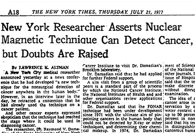
Figure 20-21a:
Headline in the New York Times, 21 July 1977: The article by Lawrence K. Altman.
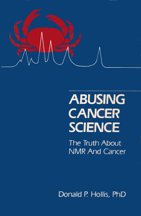
Figure 20-21b:
The cover of Donald Hollis’ book about Damadian’s scientific scam [⇒ Hollis 1987].
The attribution that Damadian made the first proposal for an MR imaging device, repeated time and again, is historically not correct.
Ian Young wrote about Damadian’s claims [⇒ Young 2004]:
“As is now well known, a huge variety of pathologic processes result in increases in the relaxation time constants, while some classes of tumor have shorter time constants than the normal tissues from which they have developed. Sadly, the many attempts that were made to correlate pathology and relaxation behavior have yielded none of the precise numerical relationships that were hoped for …
“Raymond Damadian also produced a sketch of a possible NMR imaging system … The method was, unfortunately for Damadian, one of those classic blind alleys that lead precisely nowhere.
“Donald Hollis, in his book Abusing Cancer Science: The Truth About NMR and Cancer, published in 1987, has a great deal more to say, little of it complimentary to Damadian, about the various claims he has made about both cancer diagnosis and imaging.” (Figure 20-21b).
Still, Damadian was, as it happens so often in the history of inventions, one of the many who prepared the ground — even if his published results were conclusively disproved.
Flow measurements by NMR date back as far as 1951 when the first experiment using continuous wave (CW) NMR was described by Suryan (Figure 20-22) [⇒ Suryan 1951].
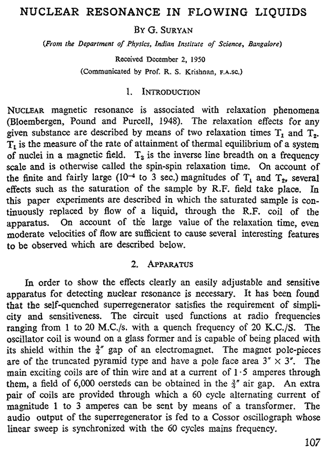
Figure 20-22:
The first page of G. Suryan’s article in the Proceedings of the Indian Academy of Sciences, 1951.
By 1959, Jerome R. Singer (Figure 20-23) had studied blood flow by NMR relaxation time measurements of blood in living humans [⇒ Singer 1959].
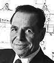 Figure 20-23: Jerome R. Singer (1921-2019).
Such measurements were not introduced into common medical practice until the mid-1980s, although patents for similar ideas were filed earlier, for instance for an NMR machine to measure blood flow in the human body by Alexander Ganssen in early 1967 [⇒ Ganssen 1967].
This machine was meant to measure the NMR signal of flowing blood at different locations of a vessel with a series of small coils, allowing to calculate the blood flow within that vessel. It could be described as an MR scanner (Figure 20-20a).
However, it was no MR imaging machine. Actual flow imaging only became possible with Lauterbur’s method.


|

|
|---|