


 he development of magnetic resonance imaging as a medical imaging technique opened the gates for several specialized imaging methods, highlighting different physical and chemical processes in the body — among them diffusion and neuronal activation. Both have become centers of attention in research.
Diffusion magnetic resonance imaging exploits the random diffusional motion of water molecules and is used in MR neuroimaging. During the recent years, additional research of numerous groups has turned this topic into one of the favorite research fields in MR imaging.
 Figure 20-53: Edward O. Stejskal (1932-2011).
Some of the essential and fundamental work was published already in 1965 by Edward O. Stejskal (Figure 20-53) and John E. Tanner (1930-2023) at the University of Wisconsin [⇒ Stejskal 1965] (cf. also Chapter 11).
Functional brain imaging (fMRI) became similarly attractive to interdisciplinary research and to groups far outside conventional science.
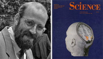
Figure 20-54:
John W. Belliveau (1959-2014) and the cover page of Science (1 November 1991) featuring one of Belliveau's first functional images through the brain.
Several research groups targeted neuronal activation, among them John W. Belliveau (Figure 20-54) and his collaborators from Boston [⇒ Belliveau 1990; 1991] who observed the enhancement of stimulated regions of the human visual cortex by following the first pass of a contrast agent through the brain.
Other groups, however, tried to exploit the differences in the magnetic properties of blood depending on its oxygenation state. The basis for BOLD-contrast was described by Pauling and Coryell in 1936 [⇒ Pauling 1936]:
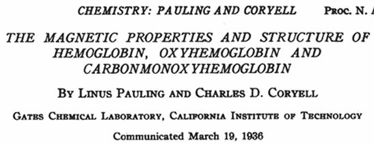
Figure 20-55:
Pauling and Coryell paper (title page).
"Over ninety years ago, on November 8, 1845, Michael Faraday investigated the magnetic properties of dried blood and made a note 'Must try recent fluid blood.' If he had determined the magnetic susceptibilities of arterial and venous blood, he would have found them to differ by a large amount (as much as twenty per cent for completely oxygenated and completely deoxygenated blood); this discovery without doubt would have excited much interest and would have influenced appreciably the course of research on blood and hemoglobin. (Figure 20-55)."
 At AT&T Bell Laboratories in New Jersey, Seiji Ogawa (Figure 20-56) and his collaborators compiled and elaborated upon these results and developed the idea of monitoring brain activity after stimulation with BOLD (Blood Oxygenation Level Dependent) contrast [⇒ Ogawa 1990].
 Figure 20-56: Seiji Ogawa.
In the race (if there was a race) of who published the first BOLD image in an animal Ogawa came in first in 1990, Robert Turner second in 1991 [⇒ Turner 1991], and third K.K. Kwong, John Belliveau and collaborators in 1992; however, they published the first human image [⇒ Kwong 1992]. Kwong had successfully performed the first human experiment in May 1991, but unfortunately his paper had been rejected by Science.
Functional MRI using BOLD is now employed as one of the principal research techniques to map the visual, auditory and sensory regions for research in neurobiology and psychology. However, its validity is still very controversial [⇒ Kim, Ogawa 2012].
The concept of using paramagnetic contrast agents to enhance pathologies was described by Paul C. Lauterbur, Maria Helena Mendonça-Dias and Andrew M. Rudin in 1978 (Figure 20-57) [⇒ Lauterbur 1978]. After injecting a manganese salt solution as contrast agent they imaged five dogs with myocardial infarctions and were able to highlight them.
Figure 20-57:
Excerpt from Lauterbur's 1978 paper in "Frontiers of Biological Energetics" on the use of manganese to highlight myocardial infarctions in dogs; the first description of the use of contrast agents in MR imaging.
In October 1983 Lauterbur's group published a major overview of paramagnetic contrast agents in MR imaging, explaining problems and questions involved in the development of possible contrast agents [⇒ Mendonça-Dias 1983].
In the period between the publication of these two articles, scientists in academia and industry took note and started their own research in the field. Several patents were taken and influenced the further progression of a hectic race, partly shrouded in secrecy.
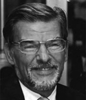 Figure 20-58: Hanns-Joachim Weinmann (1949-2009).
The former Schering company submitted a patent application for Gd-DTPA dimeglumine in July 1981 in a project involving Hanns-Joachim Weinmann (Figure 20-58) and Ulrich Speck [⇒ Weinmann 1983]. In 1984, Dennis H. Carr from London and Wolfgang Schörner from Berlin published the first images in humans [⇒ Carr DH 1984; ⇒ Schörner 1984]. In 1988, Gd-DTPA (Magnevist) became commercially available, followed shortly afterwards in 1989 by Gd-DOTA (Dotarem) from Guerbet in Paris. For a long time Dotarem was the only safe product on the market. Magnevist and several others had to be withdrawn because of side effects.
More agents entered the marketplace in the 1990s and the early 2000s.
During the last decades, more than 1000 potential MR contrast agents have been described in the literature or been patented, but only a dozen are available on the market for clinical use.


|

|
|---|